
When a child is told that a cochlear implant may help, the next message is often a wall of unfamiliar words: CT, MRI, sedation, reports. I am Dr. Shree Rao, and parents ask me the same thing every week: why two scans, and what are they looking for? A cochlear implant ct scan mri workup is not routine paperwork. Each scan answers a different question, and together they help the surgical team plan a safer operation.
What CT shows: bony anatomy, ossification, and surgical landmarks
A high-resolution CT scan of the temporal bone, the part of the skull that houses the ear, is the clearest way to see bone. It outlines the cochlea, the snail-shaped hearing organ, and the middle ear around it. It also shows the path of the facial nerve, which runs close to where the surgeon works, and the landmarks used to reach the inner ear safely.
CT can reveal inner ear malformations, signs of earlier middle ear infection, and ossification, where the fluid space inside the cochlea slowly turns to bone, often after infections such as meningitis. Ossification is not an automatic barrier to an implant, but the surgeon needs to know about it beforehand. Readers curious about how imaging supports other operations can read about how imaging guides ear surgery.
What MRI shows: the cochlear nerve, fluid spaces, and the brain
MRI uses magnets instead of X-rays and is better at soft tissue. It shows whether the cochlear nerve, the cable that carries sound signals from the ear to the brain, is present and of normal size. An implant depends on that nerve. MRI also shows whether the fluid spaces of the cochlea are open, can detect early fibrosis (scarring that may come before ossification), and images the brain, which can point to causes of hearing loss beyond the ear.
Studies generally find MRI stronger for the nerve and CT stronger for middle ear disease, and many centres recommend both. Most are small or single-centre. One review of 207 implant patients found significant abnormalities uncommon, and neither scan reliably caught every blocked cochlea, so scans are read alongside hearing tests.
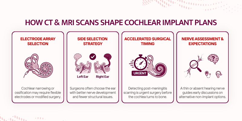
How cochlear implant ct scan mri findings change the plan: electrode choice, side selection, timing
A narrowed or partly ossified cochlea may lead the surgeon to choose a more flexible electrode array, the thin wire that sits inside the cochlea, or to plan a different surgical route. If one ear shows better nerve development or fewer obstacles, that side is often considered first.
Timing can change too. If early fibrosis is seen after meningitis, the team may move sooner, since ossification can progress with delay. If the cochlear nerve looks very thin or absent, Dr. Shree Rao discusses alternatives and sets honest expectations. Families preparing for implant surgery should know these conversations happen well before the operation.
Preparing your child for scans: sedation, timing, and what to carry
A CT scan takes only a few minutes, but the child must lie still. MRI takes longer and is noisy, so many young children need sedation or a light general anaesthetic, while some sleep naturally after a feed. The team will explain how long your child should avoid food and milk. CT involves a small dose of radiation, kept as low as reasonably possible in children and weighed against the value of a precise plan.
Plan the slot around nap time, and leave early, since Hyderabad’s monsoon traffic can delay appointments. Carry earlier hearing test reports, previous scans, records of meningitis or ear infections, and any government scheme paperwork. In multilingual homes, tell the staff which language soothes your child, whether Telugu, Hindi, or Urdu.
Conclusion
A cochlear implant ct scan mri workup gives the surgeon a map of bone, nerve, and cochlea before the first incision. CT shows the bone, MRI shows the nerve and fluid spaces, and together they guide electrode choice, side, and timing. If you are unsure which scans your child needs, you can book a consultation with Dr. Shree Rao for guidance on your next step.
FAQs
Can the MRI be skipped if the CT looks normal?
Usually not, because CT cannot show the cochlear nerve or early scarring inside the cochlea. Some centres rely on MRI alone in selected cases, so the surgeon decides.
Is MRI safe after a cochlear implant is fitted?
It depends on the implant model. Many modern implants are approved for MRI under set conditions. Keep the implant identification card handy and tell the radiology team before any scan.
How do I choose a surgeon to review my child's scans?
Look for a surgeon who studies both scans personally, explains them simply, and works alongside an audiologist and speech therapist. Families searching for the best cochlear implant specialist in India will find it useful to ask these same questions at every consultation.